Description
Number of pages 124, NEUW Publishing, ISBN 978-80-903322-4-2, Publishing Triton 978–X–80-72-54-845
Weight 0.3 kg, Dimensions 13 x 20.5 cm
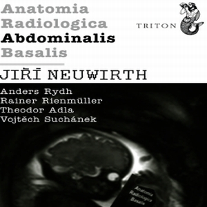
Anatomia Radiologica abdominal basalis is the third part of the paperback edition of the 4 books Anatomia Radiologica Basalis. This paperback edition is designed for medical students and doctors residents, but will also be useful for the radiologists and other professionals in their daily practice.
Anatomia Radiologica AbdominalisBasalis provides basic knowledge of the anatomy of the digestive tract and organs and blood vessels of the abdomen and pelvis on images of various imaging methods.
All 4 parts Anatomia Radiologica basalis contain 312 images of more than 4,800 identified anatomical structures and organs.
The book on images of various imaging (MRI, CT, ultrasound, radiography) described 917 unique anatomical structure of the femur after stapes and the frontal lobe after ulnar nerve.
Each book contains an index in Latin and English. For all anatomical structures in the book consistently used the Latin nomenclature according to the terminology Anatomica (1998). English description of the types of imaging techniques and projections are given in headers and footers on each side.
Complementing the book is its electronic version on the website www.anatrad.com. This electronic database is always available to owners of books and enable intuitive and simple search of anatomical structures through their on-line database on web.


